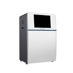


Chemiluminescence Imaging System LCIS-A23
Chemiluminescence Imaging System LCIS-A23 is a new generation, an integrated device with 10.1-inch touch screen display, for used for imaging Chemiluminescence Gel with exceptionally high quantum efficiency CCD Camera for greater sensitivity and dynamic range. It offers a 12.49 × 9.99 mm CCD Sensor Size, with 6.05 MP resolution, F0.95 motorized lens, and -30˚C cooling temperature. The 16-bit (65536 Grey Scales) CCD camera reduces the background noise during a long exposure time. The device can detect far weaker signals, offers higher sensitivity and a wider linear range. The device configuration offered as documentation of RNA, DNA, Protein, and Chemiluminescence imaging system.
| CCD Sensor Size | 12.49 × 9.99 mm |
| Resolution | 6.05 Megapixels, 2750 × 2200 |
| Pixel Density | 16bit (65536 Grey Scales) |
| Pixel Size | 4.54 × 4.54 µm |
| Lens | F0.95 motorized lens |
| Light Source | LED Epi-white light × 2 UV-Transilluminator (302 nm) White-LED Transilluminator |
| Display | 10.1 inch touch Screen Display |
| Quantum efficiency | ≥ 75 % |
| Readout noise | < 5.5e- RMS |
| Dark current | 0.0003 e/p/s |
| Dynamic range | 4 order of magnitude |
| Standard Filter | 590 nm filter |
| Filter Wheel | 8 Sockets Filter Wheel |
| Max Image Area | 260 × 210 mm |
| Cooling Temperature | -30˚C |
| Software | Image acquisition and analysis software |
| Dimension | 560 × 480 × 780 mm + 380 × 350 × 490 mm |
| Gross Weight | 39 kg |
- 10.1-inch touch screen display with user-friendly operation mode
- ≥ 75 % High Quantum efficiency of the CCD camera
- CCD Sensor Size offered is 12.49 × 9.99 mm, with a 6.05 MP resolution
- Offers convenient image navigation and browsing
- Optional attachment of different fluorescent light sources and filters
- Automatic pixel binning technology
- Marker image capture and composition with objective bands
- Small footprint - takes up minimal bench space
- Configured for maximum sensitivity to ensure even the faintest band on a blot can be captured
- Optional full configuration system Analysis System (DNA, RNA, Protein, Chemiluminescence, Fluorescence)
Offers one touch image acquisition and save
Marker image capture and composition with objective bands
Automatic access of capture parameters and calculation of the density and the peak value of each band
Convenient image navigation and browsing
It adopts image rotation, cropping and counter color processing
Advanced pixel binning technology
Optional save of analysis results as excels files
Calculation of the optical density for the quantitative analysis
It offers optimization of the visual effects by wiping background mode